TEMs have a maximum magnification of around 1000000 but images. These fine mesh grids are carefully checked.

Pin Op Bees
The first crude electron microscope was capable of magnifying objects 400 times.

Magnification of transmission electron microscope. Transmission Electron Microscopy magnification. In order to get a better idea of just how small that is think of how small a cell is. AThe TEM uses light and magnification to observe details of objects.
Nasseri and Mohammadi 99 obtained individual cellulose whiskers with length L of 8728 nm and diameter d of 153 nm with an average aspect ratio Ld of whiskers obtained was 62. X 50 to X 1000000 14 Ångstom resolution LaB6 source backscattered and secondary electron detectors Gatan Digi-PEELS Electron Energy Loss Spectrometer software and off axis imaging camera Kevex Quantum 10 mm2 X-ray detector detects elements down to boron. Transmission Electron microscope - Principle Construction Working Advantages and Disadvantages Electrons are made to pass through the specimen and the image is formed on the fluorescent screen either by using the transmitted beam or by using the diffracted beam.
The scanning electron microscope SEM has a large depth of field so can be used to examine the surface structure of specimens. TEMs have a maximum magnification of around. The scanning electron microscope is used to visualise the surfaces of cells and even whole organisms.
A scanning transmission electron microscope has achieved better than 50 pm resolution in annular dark-field imaging mode and magnifications of up to about 10000000 whereas most light microscopes are limited by diffraction to about 200 nm resolution. The maximum magnification of light microscopes is usually 1500 and their maximum resolution is 200nm due to the. The transmission electron microscope TEM is used to examine thin slices or sections of cells or tissues.
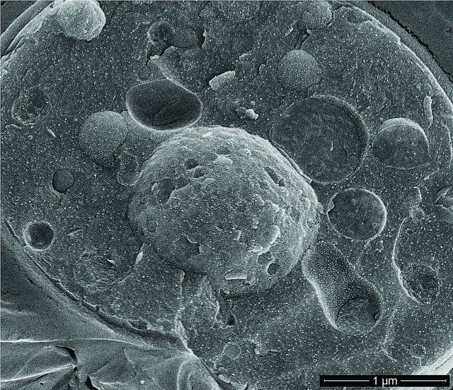
Transmission electron microscopy TEM analysis is conducted to get the actual size of the nanocrystalline cellulose fibers and in some cases the morphology. The transmission electron microscope is used to view thin specimens tissue sections molecules etc through which electrons can pass generating a projection image. A calibration sample for transmission electron microscopy TEM has been developed that performs the three major instrument calibrations for a transmission electron microscope.
A Transmission Electron Microscope TEM produces a 2D image of a thin sample and has a maximum resolution of 500000. Jiang and Hsieh obtained cellulose NFs with. Calibration Specimens for Transmission Electron Microscopy STEM Magnification Calibration Fine Mesh Grids.
Electron microscopes are normally built underground in order to reduce interference in the form of vibrations from environmental. The image magnification calibration for measurements of images the camera constant calibration for indexing diffraction patterns and the imagediffraction pattern rotation calibration for relating crystal directions. A Transmission Electron Microscope TEM utilizes energetic electrons to provide morphologic compositional and crystallographic information on samples.
The first practical electron microscope was built by in 1938and had 10 nm resolution. There are different types of Electron Microscope. The electron microscope isnow an integral part of.
Although modern electron microscopes can magnify an object 2 million times they are still based upon Ruskas prototypeand his correlation between wavelength and magnification. The transmission electron microscope TEM gives the highest magnification and resolution and it is used to observe the internal ultrastructure of cells. TEMs can magnify objects up to 2 million times.
Available as a sandwich in a folding 305 mm mesh grid or in a square mesh 25 mm. There are different types of Electron Microscope. Electron Microscopes can have magnifications of 500000.
Transmission electron microscopes TEM are microscopes that use a particle beam of electrons to visualize specimens and generate a highly-magnified image. Whereas the SEM produces images on a computer that can be colorized to enhance detailsbThe SEM is capable of magnification up to 1000000 X whereas the TEM magnifies an object up to 100000 XcThe TEM is useful for analyzing the internal organelles of a cells while the SEM gives a 3-D view of the external. The method is also known as SEM analysis and SEM microscopy and is used very effectively in microanalysis and failure analysis of.
Electron Microscopes can have magnifications of 500000. Unlike glass lenses the resolution and magnification of an electromagnetic lens is affected by changing the current through the lens whereas in a light microscope it is done by mechanically changing the lenses. They are suitable for the low magnification range of a TEM.
Scanning Electron Microscopy SEM is a test process that scans a sample with an electron beam to produce a magnified image for analysis. Where a scanning electron microscope. At a maximum potential magnification of 1 nanometer TEMs are the most powerful microscopes.
A transmission electron microscope TEM is a special type of microscope that uses electrons to create a magnified image up to 1000000x. A Transmission Electron Microscope TEM produces a 2D image of a thin sample and has a maximum resolution of 500000. What is the magnification and resolution of a light microscope.
Fewer electrons per unit area equates to a lower signal and lower signal-to-noise and if one needs or wants to severely limit the electron dose in terms of e-Å 2 there comes a point where the magnification is so high and the dose is so low that there is effectively no signal left at the detector.

Transmission Electron Microscopic Tem Image Revealed Ultrastructural Details Exhibited By Numerous Coxiella Burnetii Ba Q Fever Microscopic Images Bacteria

Pin On Viruses The Ultimate Killers

Fragment Of Surface Of Diatom Cell Magnified 11000x Microscopic Photography Geometry In Nature Macro And Micro

Transmission Electron Microscope Tem Micrograph Showing Several Peripheral Myelinated Fibers And A Sch Electron Microscope Microscopy Microscopic Photography
Yeast Cell Microscopic Photography Microscopy Electrons

Here Are Algae And Bacteria Magnified 5000 Times Scanning Electron Microscope Personal Hygiene Electron Microscope

Visualization Of The Cell Using Em Scanning Electron Microscope Microscopic Photography The Cell

Histology Gallery Eukaryotic Cell Microscopic Photography Cell Biology

Euglena Tem Stock Image C009 4026 Organic Art Bio Art Science Nerd

Trachea 8000x Magnification Scanning Electron Microscope Electron Microscope Electron Microscope Images