I TEM analysis is one of the few methods. Medical Definition of transmission electron microscope.
Sem Vs Tem Electron Microscopy Microbe Online
Transmission electron microscope TEM an electron microscope that produces highly magnified images of ultrathin tissue sections or other specimens.
Definition of transmission electron microscope. Specially prepared materials samples may. They are also the most powerful microscopic tool available to-date capable of producing high-resolution detailed images 1 nanometer in size. This increased resolution allows us to study ultrastucture of organelles viruses and macromolecules.
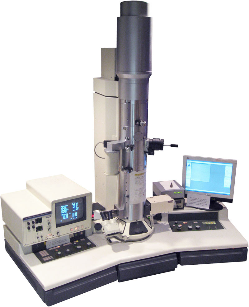
The Transmission Electron Microscope TEM Similar to the general scheme of a light microscope a transmission electron microscope 1 2 consists of an electron source a condenser system an objective lens and a projector system as shown in Fig. TEMs are costly large cumbersome instruments that require special housing and maintenance. A conventional electron microscope which produces an image of a cross-sectional slice of a specimen all points of which are illuminated by the electron beam at the same time compare scanning electron microscope.
Transmission electron microscopes TEM are microscopes that use a particle beam of electrons to visualize specimens and generate a highly-magnified image. Transmission electron microscope TEM - definition of transmission electron microscope TEM by The Free Dictionary. Transmission electron microscope noun A form of electron microscope in which an image is derived from electrons which have passed through the specimen in particular one in which the whole image is formed at once rather than by scanning.
The transmission electron microscope TEM uses a high voltage electron beam emitted by an electron gun. An optical instrument that uses a lens or a combination of lenses to produce magnified images of small objects especially of objects too small to be. It can be considered as a golden standard for the characterization of NM for several reasons 1.
Since the wavelength of an electron is much smaller than the wavelength of visible light diffraction effects occur at much smaller physical dimensions. Many transmission electron microscopes have additional instruments attached to it such as an. Microscopio electrónico de transmisión instrumento usado para visualizar las células con una capacidad superior de un millón más de visualización que el microscopio común.
1Many transmission electron microscopes have additional instruments attached to it such as an X-ray detector andor an energy loss spectrometer in order to be able to perform elemental. A Transmission Electron Microscope produces images via the interaction of electrons with a sample. Transmission electron microscope TEM type of electron microscope that has three essential systems.
An electron beam passes through the metal-impregnated specimen and is focused by magnetic lenses into an image. The transmission electron microscope TEM operates on many of the same optical principles as the light microscope. In order to get a better idea of just how small that is think of how small a cell is.
1 an electron gun which produces the electron beam and the condenser system which focuses the beam onto the object 2 the image-producing system consisting of the objective lens movable specimen stage and intermediate and projector lenses which focus the electrons passing through. Similar to the general scheme of a light microscope a transmission electron microscope 1 2 consists of an electron source a condenser system an objective lens and a projector system as shown in Fig. Transmission Electron Microscope TEM definition.
Electromagnetic lenses are used to focus the electron beam on the sample. As the electron beam passes through the sample and the atoms. What is a Transmission Electron Microscope.
Transmission electron microscope TEM an electron microscope that produces highly magnified images of ultrathin tissue sections or other specimens. The TEM is analogous in many ways to the conventional compound light microscope. English-Spanish Medical Dictionary Farlex 2012.
An electron beam passes through the metal-impregnated specimen and is focused by magnetic lenses into an image. The TEM has the added advantage of greater resolution. Definition of Transmission Electron Microscopy TEM Electron microscopy is an imaging technique that uses an electron beam to probe a material.
A transmission electron microscope is an electron microscope used to see objects far smaller than cells. Transmission electron microscopy TEM is a versatile technique to analyse the size morphology crystallographic structure and chemical composition of a wide range of nanomaterials NM. In a Transmission electron microscope the electron beam is transmitted through a very thin specimen or object and forms a highly magnified and detailed image of the sample.
TEMs can magnify objects up to 2 million times. This microscope uses electron beams instead of light. The transmission electron microscope TEM The transmission electron microscope is used to view thin specimens through which electrons can pass generating a projection image.
There are different types of Electron Microscope. An electron microscope on the other hand uses a beam of electrons rather than light to form the image.

Field Emission Scanning Electron Microscopy Fesem Photometrics
Ad Get More Out of Your Digital Microscope and Choose a DSX1000 Model that Suits Your Needs.
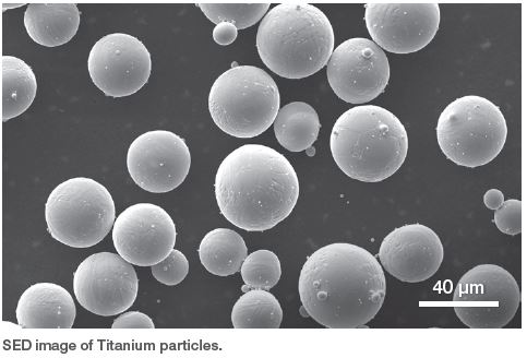
Electron microscope magnification range. To see the results look at the image below. SEM analysis provides information about both the surface topography and cross-sections. Electron microscope definition.
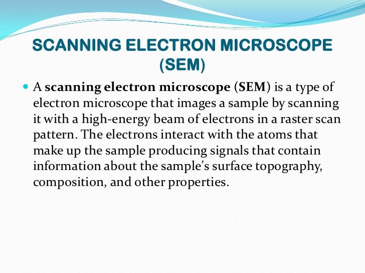
To put that into perspective the human eye can see things down to single strand of hair the thickness of which is about 0065 millimeters. The magnification of an electron microscope may be as high as 10000000x with a resolution of 50 picometers 005 nanometers. The scanning electron microscope SEM uses a focused beam of high-energy electrons to generate a variety of signals at the surface of solid specimens.
Click to see full answer. It provides information from coarse millimeter-scale particles up to real nanoparticles in a magnification range of 20200000 Yin et al 2014. It is a special type of microscope having a high resolution of images able to magnify objects in nanometres which are formed by controlled use of electrons in vacuum captured on a phosphorescent screen.
Microscope depend on the parameters of this electron beam. As a comparison total maximum magnifications are 1000000x for electron microscopes and about 1500x for a light microscope. Electron microscope magnification standard providing precise calibration in the magnification range 5000X-2000000X.
An electron microscope can achieve magnification in excess of 100000x compared with 1000X magnification with light microscopy. A Transmission Electron Microscope TEM produces a 2D image of a thin sample and has a maximum resolution of 500000. It is mainly considered that the.
A lower end floor top SEM will have magnification around x300000 while higher end SEMs can go up to x1000000. The magnification of an electron microscope may be as high as 10000000x with a resolution of 50 picometers 005 nanometers. There is a minimum magnification necessary for the detail present in an image to be resolved and this value is usually rather arbitrarily set as 500 times the numerical aperture 500 x NA and defined by the equation.
The range of useful magnification for an objectiveeyepiece combination is defined by the numerical aperture of the microscope optical system. Scanning electron microscopes have smaller maximum magnifications than transmission electron microscopes. Why is a vacuum needed in an electron microscope.
Also taking into account empty space between particles a field of view of 25-30 microns is enough for such sample. A table-top SEM can have maximum magnification of x60000 for lower end models while higher end models have maximum magnification of about x150000. A light microscope can magnify things up to 2000x but an electron microscope can magnify between 1 and 50 million times depending on which type you use.
Electron Microscopes can have magnifications of 500000. The spot size determines the resolution of the microscope as well as usable magnification at stable picture sharpness. Trust Our Long History in Cutting-Edge Optics for Superior Quality Advanced Performance.
A Transmission Electron Microscope TEM produces a 2D image of a thin sample and has a maximum resolution of 500000. TEMs have a maximum magnification of around x1000000 but images can be enlarged beyond that photographically. The signals that derive from electron-sample interactions reveal information about the sample including external morphology texture chemical composition and crystalline structure and orientation of materials making up the sample.
Trust Our Long History in Cutting-Edge Optics for Superior Quality Advanced Performance. A light microscope uses light as its illumination source where as. High-quality images Electron microscopes produce highly detailed images of structures which are of high quality revealing complex and delicate.
Magnification and higher resolution Electron microscopes provide an image resolution in the range of up to 02 nm. At a maximum potential magnification of 1 nanometer the transmission electron microscope is the most powerful microscopes for a wide range of educational science and industry applications. The magnification depends on the type and make of EM.
Larger particles will require lower magnifications in order to show multiple particles in a single image and the required magnification for such images can range from 20000x to 100000x. The resolution limit of electron microscopes is about 02nm the maximum useful magnification an electron microscope can provide is about 1000000x. Electron Microscopes can have magnifications of 500000.
On the other hand if your interest lies in the structure of a particle a close up is needed and the observed area must be closer to 2-3 microns if not smaller. As always specimen properties will dictate at least some of the magnifications used. The limit of resolution of the transmission electron microscope is now less than 1.
This makes electron microscopes more powerful than light microscopes. Typically the standard light microscope will max out at about 1500X magnification and the electron microscope will be able to achieve 200000X magnification. An electron microscope is a microscope that uses a beam of accelerated electrons as a source of illumination.
Spot size aperture angle and beam intensity. Ad Get More Out of Your Digital Microscope and Choose a DSX1000 Model that Suits Your Needs. Each type of microscope however has its limitations whether its in the optics or the way the tissues are prepared or mounted.
There are different types of Electron Microscope.
Electron source to produce electrons magnetic lenses to de-magnify the beam magnetic coils to control and modify the beam apertures to define the beam prevent electron spray etc. Two sets of condenser lenses focus the electron beam on the specimen and then into a thin tight beam.

Scanning Electron Microscope Sem Microbe Notes
The electrons are emitted from a filament and collimated into a beam in the electron source.
Working principle of scanning electron microscope. Scanning electron microscope principle working SEM - YouTube. In a scanning electron microscope the specimen is exposed to a narrow electron beam from an electron gun which rapidly moves over or scans the surface of the specimen Figure 413. When a cathode emits electrons under the application of a very high electric field it is also known as a field emitter-FE- SEM which gives better images.
At the heart of a scanning electron microscope is a high-energy electron source positioned above a series of condenser lenses and apertures which focus these electrons into a beam. Scanning electron microscope principle. A high voltage current is applied which results in the excitation of the electrons in the form of a continuous stream that is used as a beam of light.
These high speed primary electrons on falling over the sample produces low energy secondary electrons. Principles of Scanning Electron Microscopy. What is the working principle of scanning electron microscope.
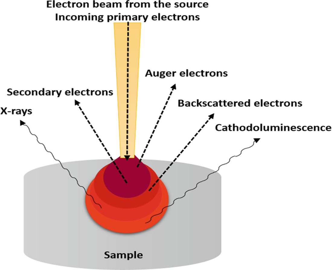
The beam is then focused on the sample surface by a set of lenses in the electron column. Illustration of several signals generated by the electron beamspecimen inter-action in the scanning electron microscope and the regions from which the signals can be detected. Electron microscopes use signals arising from the interaction of an electron beam with the sample to obtain information about structure morphology and composition.
Used to protect the electronic beam from interference with air. COMPONENTS OF ELECTRON MICROSCOPE 1. SEM equipment is similar to a television.
The electrons are emitted from a filament and collimated into a beam in the electron source. Scanning Electron Microscope functions exactly as their optical counterparts except that they use a focused beam of electrons instead of light to. The metal used in an electron microscope is tungsten.
Stream of electrons are produced by the electron gun and these primary electrons are accelerated by the grid and anode. This causes the release of a shower of secondary electrons and other types of radiations from the specimen surface. Control the final converge nce angle of the electron beam onto.
Scanning electron microscopes SEMs use an electron beam to image samples with a resolution down to the nanometer scale. Fundamentals of Scanning Electron Microscopy 3 1 Beam Backscatterred electrons Secondary electrons Auger electrons Characteristic x-rays X-ray continuum FIGURE 12. The incident electron beam is scanned in a raster pattern across the surface of the sample and the backscattered or the secondary electrons emitted are detected.
Scanning electron microscope sem works on the principle of interaction of electron beam accelerated electrons - Advertisement -. Electron optical column consists of. The scanning electron microscope SEM uses a focused beam of high-energy electrons to generate a variety of signals at the surface of solid specimens.
Principle of Scanning Electron Microscopy. With the atoms of a sample at various depths and reveal the information in the form of signals. The electron gun generates electrons.
This course represents the fundamentals on the working principles of all main components of the modern microscopes such as electron and ion optics as well as diverse signal detectors on the physics of the particle interaction with the matter and of the image formation in different operation modes. Working Principle of Electron microscope. The principle of SEM is to use a focused beam of high-energy electrons to generate a variety of signals at the surface of solid specimens such as secondary electrons backscattered electrons and X-rays.
How Scanning Electron Microscopy Works. The scanning electron microscope SEM Principle of Electron Microscope. Scanning electron microscopy SEM uses electrons for imaging.
How a scanning electron microscope SEM works. These accelerated primary electrons are made to be incident on the sample through condensing lenses and scanning coil. Control the number of electrons which reach the sample.
The lenses used in the electron microscope are magnetic coils. The signals that derive from electron-sample interactions reveal information about the sample including external morphology texture chemical composition and crystalline structure and orientation. Scanning electron microscopes SEMs use an electron beam to image samples with a resolution down to the nanometer scale.
The electron beam behaves like a wavefront with wavelength about a million times shorter than lightwaves. Transmission Electron Microscope Uses in Microscopy Advantages and Disadvantages.
![]()
Transmission Electron Microscope Instrument Britannica
Working Principle of Electron microscope.
Working principle of transmission electron microscope. Transmission electron microscope. The major difference is the light microscope uses artificial light or natural light to create an image of the specimen. In order to get a better idea of just how small that is think of how small a cell is.
The transmission electron microscope TEM The scanning electron microscope SEM Principle of Electron Microscope. TEM Transmission electron microscopy. Working principle of Transmission Electron Microscope Electron Microscope follows the same principle as a light microscope follow.
Since the wavelength o f electrons are 100000 times shorter than visible light the electron microscopes have greater resolving power. 1 Transmission Electron Microscope TEM. Scanning transmission electron microscopy STEM combines the principles of transmission electron microscopy and scanning electron microscopy and can be performed on either type of instrument.
Electrons are made to pass through the specimen and the image is formed on the fluorescent screen either by using the transmitted beam or by using the diffracted beam. Guide Scanning Electron Microscopy Working Principle 8 Transmission electron microscopy TEM In TEM the accelerated electrons pass through the specimen. Working principle of TEM.
Like TEM STEM requires very thin samples and looks primarily at beam electrons transmitted by the sample. Microscopy Transmission electron microscopy TEM an abbreviation which can also stand for the instrument a transmission electron microscope is a microsco. The metal used in an electron microscope is tungsten.
TEMs produce high-resolution two-dimensional images allowing. There are two types of electron microscopes. Transmission Electron Microscopy In a conventional transmission electron microscope a thin specimen is irradiated with an electron beam of uniform current density.
Objective lens provides the formation of either. TEMs can magnify objects up to 2 million times. The electron gun generates electrons.
The transmitted ones then become focused as an enlarged image onto a fluorescent screen which. To familiarize the technique of sample preparation for transmission electron microscopy. Transmission electron microscopy TEM is the original form of electron microscopy and analogues to the optical microscope.
A high voltage current is applied which results in the excitation of the electrons in the form of a continuous stream that is used. In this microscope an electron beam from an electron gun is transmitted through an ultra-thin section of the microscopic object and the image is magnified by the electromagnetic fields. Electrons are made to pass through the specimen and the image is formed on the fluorescent screen either by using the transmitted beam or by using the diffracted beam.
TEM functions under the principle of optical microscopy. Transmission electron microscopes TEM are microscopes that use a particle beam of electrons to visualize specimens and generate a highly-magnified image. Under vacuum conditions the electron beam is accelerated by high pressure to form scattering electrons and transmission.
Electrons are emitted from the electron gun and illuminate the specimen through a two or three stage condenser lens system. Transmission electron microscope. At a maximum potential magnification of 1 nanometer TEMs are the most powerful microscopes.
A Transmission Electron Microscope TEM utilizes energetic electrons to provide morphologic compositional and crystallographic information on samples. The electrons are replaced by photons glass lenses are replaced by electromagnetic lenses and images are viewed in a screen instead of an eyepiece. Transmission electron microscopy principle and working lecture TEM - This microscopy lecture is going to explain the Transmission electron microscopy princ.
Electron microscopes use signals arising from the interaction of an electron beam with the sample to obtain information about structure morphology and composition. What is a Transmission Electron Microscope. Transmission electron microscopy uses high energy electrons up to 300 kV accelerating voltage which are accelerated to nearly the speed of light.
Electron beams are used in electron microscope to illuminate the specimen and thus creates an image. A transmission electron microscope TEM is a special type of microscope that uses electrons to create a magnified image up to a million times. It can achieve a resolution.
Transmission electron microscope that is transmission electron microscope is usually called electron microscope or electron microscope EM is the most widely used class of electron microscope. Two sets of condenser lenses focus the electron beam on the specimen and then into a thin tight beam.
Furthermore you will discover how energy-dispersive x-ray spectroscopy can be paired with scanning electron microscopy to gain elemental information about samples. The Scanning Electron Microscope A Small World of Huge Possibilities.

Pin On A Luna Blue Stock Video
Today I finally produced an image with my DIY scanning electron microscope.

Scanning electron microscope video. You will also be able to explain the benefits of environmental scanning electron microscopy. Objectives of the course are to define and illustrate the major components of the SEM as well as describe methodology of. She explains how a scanning electron microscope works by knocking electrons off the surface of a sample.
What is Scanning Electron Microscopy SEM A typical SEM instrument showing the electron column sample chamber EDS detector electronics console and visual display monitors. Scanning electron microscope column 1. Welcome to the exciting world of scanning electron microscope photography and video.
Understand more about SEM microscopy in Why we use SEM Artefacts in the SEM and Sputter coating. A scanning electron microscope SEM is a type of microscope that relies on tiny particles called electrons instead of light in order to generate an image. Scanning Electron Microscope- Main components- Basic principle- Practical procedure- Imaging of surfaces and chemical analysisResponsible for this video.
HIV Virus 004 3D Rendering. 4K and HD video ready for any NLE immediately. Its been 50 years since Cambridge Scientific Instruments launched the first commercial scanning electron microscope SEM in 1965.
There is still a lot of work left to do in making the image higher resolution and eliminating sources of noise however. Getty Images offers exclusive rights-ready and premium royalty-free analog HD and 4K video of the highest quality. Ive spent the last few months working on this project and am encouraged by todays success.
This short film is about SuperSTEM which is the UKs national facility for aberration-corrected scanning transmission electron microscopy STEM. DIY scanning electron microscope - Overview video. Get a 25000 second scanning electron microscope image of stock footage at 2398fps.
Electrons are primarily accelerated toward an anode that. Researchers perception of the nanoscale world has never been the same. The course is designed as an introduction to the SEM and as a research tool for students who have had no previous SEM experience.
Explore subjects from the ordinary to the extraordinary--from common objects and creatures to exotic animals and plants microbiology and high technology. Choose from a wide range of similar scenes. Download this video clip and other motion backgrounds special effects After Effects templates and more.
View All Photo Categories. Find professional Scanning Electron Microscope videos and stock footage available for license in film television advertising and corporate uses. Therefore the first part of this paper discusses fundamentals of scanning electron microscopy with particular emphasis on signal generation caused by primary electron beam interaction with the object collection and.
Located at the top of the column where free electrons are generated by thermionic emission from a tungsten filament at 2700K. The scanning electron microscope SEM uses a focused beam of high-energy electrons to generate a variety of signals at the surface of solid specimens. The Scanning Electron Microscope SEM at Solar Atmospheres with examples of the SEM value in detecting material characterizations.
Video clip id 1019787301. Find your scanning electron microscope easily amongst the 32 products from the leading brands Hitachi Zeiss RENISHAW on DirectIndustry the industry specialist for your professional purchases. This collection of photographic works spans 1974 to the present.
Royalty free stock video and stock footage. The scanning electron microscope is a specialized closed-circuit television system which must be understood before the properties of thin films can be monitored using it. Scanning electron microscope SEM.
The filament is inside the Wehnelt which controls the number of electrons leaving the gun. This electron microscopy lecture explains about the Scanning electron microscopy or SEM principle and advantagesSEM stands for scanning electron microscope. Liz Girvan is a SEM expert with Otago Micro and Nanoscale Imaging.
Scanning electron microscope image of the HIV virus.