Electron source to produce electrons magnetic lenses to de-magnify the beam magnetic coils to control and modify the beam apertures to define the beam prevent electron spray etc. Two sets of condenser lenses focus the electron beam on the specimen and then into a thin tight beam.

Scanning Electron Microscope Sem Microbe Notes
The electrons are emitted from a filament and collimated into a beam in the electron source.
Working principle of scanning electron microscope. Scanning electron microscope principle working SEM - YouTube. In a scanning electron microscope the specimen is exposed to a narrow electron beam from an electron gun which rapidly moves over or scans the surface of the specimen Figure 413. When a cathode emits electrons under the application of a very high electric field it is also known as a field emitter-FE- SEM which gives better images.
At the heart of a scanning electron microscope is a high-energy electron source positioned above a series of condenser lenses and apertures which focus these electrons into a beam. Scanning electron microscope principle. A high voltage current is applied which results in the excitation of the electrons in the form of a continuous stream that is used as a beam of light.
These high speed primary electrons on falling over the sample produces low energy secondary electrons. Principles of Scanning Electron Microscopy. What is the working principle of scanning electron microscope.
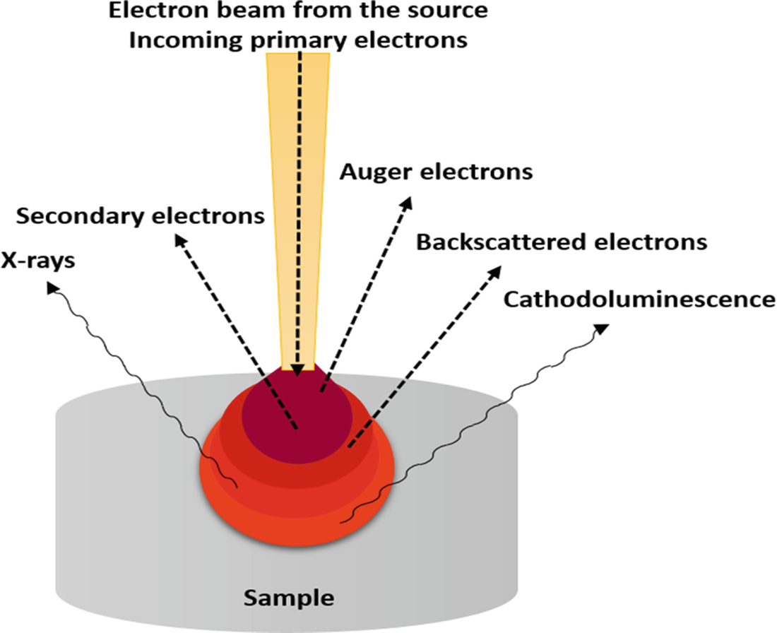
The beam is then focused on the sample surface by a set of lenses in the electron column. Illustration of several signals generated by the electron beamspecimen inter-action in the scanning electron microscope and the regions from which the signals can be detected. Electron microscopes use signals arising from the interaction of an electron beam with the sample to obtain information about structure morphology and composition.
Used to protect the electronic beam from interference with air. COMPONENTS OF ELECTRON MICROSCOPE 1. SEM equipment is similar to a television.
The electrons are emitted from a filament and collimated into a beam in the electron source. Scanning Electron Microscope functions exactly as their optical counterparts except that they use a focused beam of electrons instead of light to. The metal used in an electron microscope is tungsten.
Stream of electrons are produced by the electron gun and these primary electrons are accelerated by the grid and anode. This causes the release of a shower of secondary electrons and other types of radiations from the specimen surface. Control the final converge nce angle of the electron beam onto.
Scanning electron microscopes SEMs use an electron beam to image samples with a resolution down to the nanometer scale. Fundamentals of Scanning Electron Microscopy 3 1 Beam Backscatterred electrons Secondary electrons Auger electrons Characteristic x-rays X-ray continuum FIGURE 12. The incident electron beam is scanned in a raster pattern across the surface of the sample and the backscattered or the secondary electrons emitted are detected.
Scanning electron microscope sem works on the principle of interaction of electron beam accelerated electrons - Advertisement -. Electron optical column consists of. The scanning electron microscope SEM uses a focused beam of high-energy electrons to generate a variety of signals at the surface of solid specimens.
Principle of Scanning Electron Microscopy. With the atoms of a sample at various depths and reveal the information in the form of signals. The electron gun generates electrons.
This course represents the fundamentals on the working principles of all main components of the modern microscopes such as electron and ion optics as well as diverse signal detectors on the physics of the particle interaction with the matter and of the image formation in different operation modes. Working Principle of Electron microscope. The principle of SEM is to use a focused beam of high-energy electrons to generate a variety of signals at the surface of solid specimens such as secondary electrons backscattered electrons and X-rays.
How Scanning Electron Microscopy Works. The scanning electron microscope SEM Principle of Electron Microscope. Scanning electron microscopy SEM uses electrons for imaging.
How a scanning electron microscope SEM works. These accelerated primary electrons are made to be incident on the sample through condensing lenses and scanning coil. Control the number of electrons which reach the sample.
The lenses used in the electron microscope are magnetic coils. The signals that derive from electron-sample interactions reveal information about the sample including external morphology texture chemical composition and crystalline structure and orientation. Scanning electron microscopes SEMs use an electron beam to image samples with a resolution down to the nanometer scale.