TEMs have a maximum magnification of around 1000000 but images. These fine mesh grids are carefully checked.

Pin Op Bees
The first crude electron microscope was capable of magnifying objects 400 times.

Magnification of transmission electron microscope. Transmission Electron Microscopy magnification. In order to get a better idea of just how small that is think of how small a cell is. AThe TEM uses light and magnification to observe details of objects.
Nasseri and Mohammadi 99 obtained individual cellulose whiskers with length L of 8728 nm and diameter d of 153 nm with an average aspect ratio Ld of whiskers obtained was 62. X 50 to X 1000000 14 Ångstom resolution LaB6 source backscattered and secondary electron detectors Gatan Digi-PEELS Electron Energy Loss Spectrometer software and off axis imaging camera Kevex Quantum 10 mm2 X-ray detector detects elements down to boron. Transmission Electron microscope - Principle Construction Working Advantages and Disadvantages Electrons are made to pass through the specimen and the image is formed on the fluorescent screen either by using the transmitted beam or by using the diffracted beam.
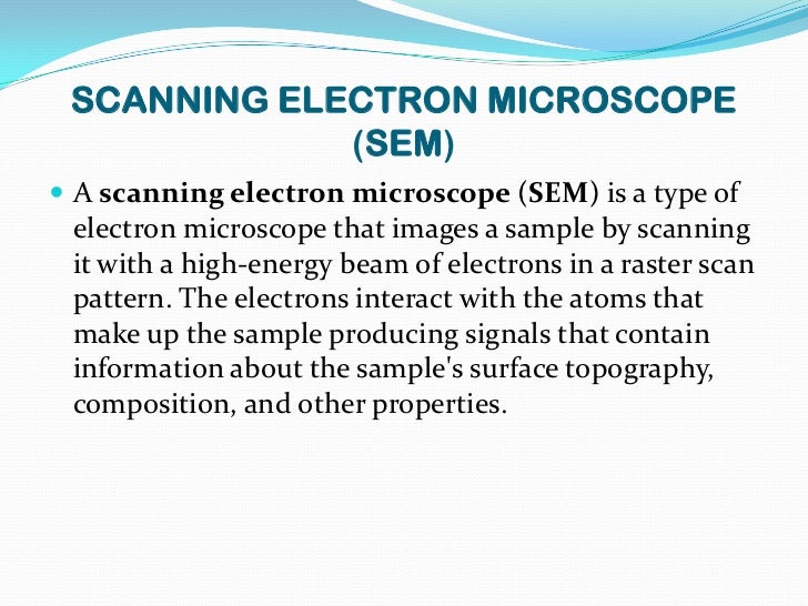
The scanning electron microscope SEM has a large depth of field so can be used to examine the surface structure of specimens. TEMs have a maximum magnification of around. The scanning electron microscope is used to visualise the surfaces of cells and even whole organisms.
A scanning transmission electron microscope has achieved better than 50 pm resolution in annular dark-field imaging mode and magnifications of up to about 10000000 whereas most light microscopes are limited by diffraction to about 200 nm resolution. The maximum magnification of light microscopes is usually 1500 and their maximum resolution is 200nm due to the. The transmission electron microscope TEM is used to examine thin slices or sections of cells or tissues.
Transmission electron microscopy TEM analysis is conducted to get the actual size of the nanocrystalline cellulose fibers and in some cases the morphology. The transmission electron microscope is used to view thin specimens tissue sections molecules etc through which electrons can pass generating a projection image. A calibration sample for transmission electron microscopy TEM has been developed that performs the three major instrument calibrations for a transmission electron microscope.
A Transmission Electron Microscope TEM produces a 2D image of a thin sample and has a maximum resolution of 500000. Jiang and Hsieh obtained cellulose NFs with. Calibration Specimens for Transmission Electron Microscopy STEM Magnification Calibration Fine Mesh Grids.
Electron microscopes are normally built underground in order to reduce interference in the form of vibrations from environmental. The image magnification calibration for measurements of images the camera constant calibration for indexing diffraction patterns and the imagediffraction pattern rotation calibration for relating crystal directions. A Transmission Electron Microscope TEM utilizes energetic electrons to provide morphologic compositional and crystallographic information on samples.
The first practical electron microscope was built by in 1938and had 10 nm resolution. There are different types of Electron Microscope. The electron microscope isnow an integral part of.
Although modern electron microscopes can magnify an object 2 million times they are still based upon Ruskas prototypeand his correlation between wavelength and magnification. The transmission electron microscope TEM gives the highest magnification and resolution and it is used to observe the internal ultrastructure of cells. TEMs can magnify objects up to 2 million times.
Available as a sandwich in a folding 305 mm mesh grid or in a square mesh 25 mm. There are different types of Electron Microscope. Electron Microscopes can have magnifications of 500000.
Transmission electron microscopes TEM are microscopes that use a particle beam of electrons to visualize specimens and generate a highly-magnified image. Whereas the SEM produces images on a computer that can be colorized to enhance detailsbThe SEM is capable of magnification up to 1000000 X whereas the TEM magnifies an object up to 100000 XcThe TEM is useful for analyzing the internal organelles of a cells while the SEM gives a 3-D view of the external. The method is also known as SEM analysis and SEM microscopy and is used very effectively in microanalysis and failure analysis of.
Electron Microscopes can have magnifications of 500000. Unlike glass lenses the resolution and magnification of an electromagnetic lens is affected by changing the current through the lens whereas in a light microscope it is done by mechanically changing the lenses. They are suitable for the low magnification range of a TEM.
Scanning Electron Microscopy SEM is a test process that scans a sample with an electron beam to produce a magnified image for analysis. Where a scanning electron microscope. At a maximum potential magnification of 1 nanometer TEMs are the most powerful microscopes.
A transmission electron microscope TEM is a special type of microscope that uses electrons to create a magnified image up to 1000000x. A Transmission Electron Microscope TEM produces a 2D image of a thin sample and has a maximum resolution of 500000. What is the magnification and resolution of a light microscope.
Fewer electrons per unit area equates to a lower signal and lower signal-to-noise and if one needs or wants to severely limit the electron dose in terms of e-Å 2 there comes a point where the magnification is so high and the dose is so low that there is effectively no signal left at the detector.
I TEM analysis is one of the few methods. Medical Definition of transmission electron microscope.
Sem Vs Tem Electron Microscopy Microbe Online
Transmission electron microscope TEM an electron microscope that produces highly magnified images of ultrathin tissue sections or other specimens.
Definition of transmission electron microscope. Specially prepared materials samples may. They are also the most powerful microscopic tool available to-date capable of producing high-resolution detailed images 1 nanometer in size. This increased resolution allows us to study ultrastucture of organelles viruses and macromolecules.
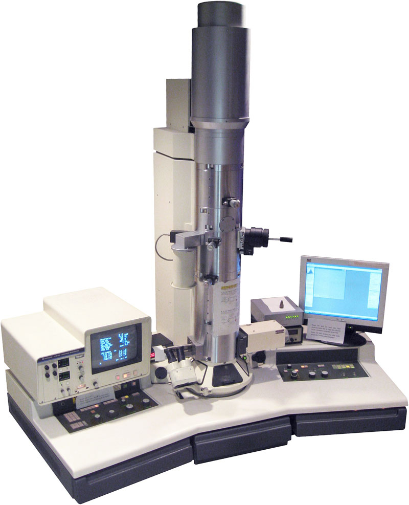
The Transmission Electron Microscope TEM Similar to the general scheme of a light microscope a transmission electron microscope 1 2 consists of an electron source a condenser system an objective lens and a projector system as shown in Fig. TEMs are costly large cumbersome instruments that require special housing and maintenance. A conventional electron microscope which produces an image of a cross-sectional slice of a specimen all points of which are illuminated by the electron beam at the same time compare scanning electron microscope.
Transmission electron microscopes TEM are microscopes that use a particle beam of electrons to visualize specimens and generate a highly-magnified image. Transmission electron microscope TEM - definition of transmission electron microscope TEM by The Free Dictionary. Transmission electron microscope noun A form of electron microscope in which an image is derived from electrons which have passed through the specimen in particular one in which the whole image is formed at once rather than by scanning.
The transmission electron microscope TEM uses a high voltage electron beam emitted by an electron gun. An optical instrument that uses a lens or a combination of lenses to produce magnified images of small objects especially of objects too small to be. It can be considered as a golden standard for the characterization of NM for several reasons 1.
Since the wavelength of an electron is much smaller than the wavelength of visible light diffraction effects occur at much smaller physical dimensions. Many transmission electron microscopes have additional instruments attached to it such as an. Microscopio electrónico de transmisión instrumento usado para visualizar las células con una capacidad superior de un millón más de visualización que el microscopio común.
1Many transmission electron microscopes have additional instruments attached to it such as an X-ray detector andor an energy loss spectrometer in order to be able to perform elemental. A Transmission Electron Microscope produces images via the interaction of electrons with a sample. Transmission electron microscope TEM type of electron microscope that has three essential systems.
An electron beam passes through the metal-impregnated specimen and is focused by magnetic lenses into an image. The transmission electron microscope TEM operates on many of the same optical principles as the light microscope. In order to get a better idea of just how small that is think of how small a cell is.
1 an electron gun which produces the electron beam and the condenser system which focuses the beam onto the object 2 the image-producing system consisting of the objective lens movable specimen stage and intermediate and projector lenses which focus the electrons passing through. Similar to the general scheme of a light microscope a transmission electron microscope 1 2 consists of an electron source a condenser system an objective lens and a projector system as shown in Fig. Transmission Electron Microscope TEM definition.
Electromagnetic lenses are used to focus the electron beam on the sample. As the electron beam passes through the sample and the atoms. What is a Transmission Electron Microscope.
Transmission electron microscope TEM an electron microscope that produces highly magnified images of ultrathin tissue sections or other specimens. The TEM is analogous in many ways to the conventional compound light microscope. English-Spanish Medical Dictionary Farlex 2012.
An electron beam passes through the metal-impregnated specimen and is focused by magnetic lenses into an image. The TEM has the added advantage of greater resolution. Definition of Transmission Electron Microscopy TEM Electron microscopy is an imaging technique that uses an electron beam to probe a material.
A transmission electron microscope is an electron microscope used to see objects far smaller than cells. Transmission electron microscopy TEM is a versatile technique to analyse the size morphology crystallographic structure and chemical composition of a wide range of nanomaterials NM. In a Transmission electron microscope the electron beam is transmitted through a very thin specimen or object and forms a highly magnified and detailed image of the sample.
TEMs can magnify objects up to 2 million times. This microscope uses electron beams instead of light. The transmission electron microscope TEM The transmission electron microscope is used to view thin specimens through which electrons can pass generating a projection image.
Transmission electron microscope TEM and scanning electron microscope SEM work on the same basic principle. Electron gun is a heated tungsten filament which emitted electron beams.

Visualization Of The Cell Using Em Scanning Electron Microscope Microscopic Photography The Cell
What Is An Electron Microscope Definition Types Uses Study Com.

Parts of transmission electron microscope. The beam of electrons are the focused on the specimen by the. Electron microscope definition. By connecting this gun to a high-voltage source of about 100 300 kV the gun begins to emit electrons by either thermionic or field electron emission.
Instead of detecting electrons being transmitted from an electron source Scanning Electron Microscopy uses the primary electron beam to excite the specimen. Electron Microscopes contain these following parts. Transmission electron microscope TEM type of electron microscope that has three essential systems.
Electron source Electromagnetic lens system Sample holder Imaging system The electron source is an electron gun which consists of a tungsten filament. The TEM consists of an electron emission source which may be a tungsten filament or a lanthanum hexaboride LaB 6 source known as electron gun. DIFFERENCE BETWEEN OPTICAL MICROSCOPE AND ELECTRON MICROSCOPE.
It is the source of electrons. Air needs to be pumped out of the vacuum chamber creating a space where electrons are able to move. The organization of the transmission electron microscope TEM is similar to that of the light microscope.
An electron microscope consists of an electric gun microscope column electromagnetic coils a fluorescent screen and some other accessories described below. Transmission electron microscopes TEM are microscopes that use a particle beam of electrons to visualize specimens and generate a highly-magnified image. Simple Microscope Parts Functions Diagram And Labelling.
Scanning-transmission electron microscopes irradiate the sample in a sequential raster pattern like scanning electron microscopes but still form images from those electrons that are transmitted through the specimen ie the electron detector is on the far side of the specimen unlike the case for scanning electron microscopes. Electron Microscope is scientific instrument that use a beam of highly energetic electrons to examine objects on a very fine scale ODUCTION. This filament emits electrons when it is heated.
It is a special type of microscope having a high resolution of images able to magnify objects in nanometres which are formed by controlled use of electrons in vacuum captured on a phosphorescent screen. It is usually a hairpin-shaped tungsten wire. 1 an electron gun which produces the electron beam and the condenser system which focuses the beam onto the object 2 the image-producing system consisting of the objective lens movable specimen stage and intermediate and projector lenses which focus the electrons passing through the.
Parts of A Transmission Electron Microscope 1. A Transmission Electron Microscope produces a high-resolution black and white image from the interaction that takes place between prepared samples and energetic electrons in the vacuum chamber. The illumination source or electron gun in a thermo-ionic emission TEM works much like a light bulb.
A TEM is composed of the following. Simple Microscope Parts Functions Diagram And Labelling. EM consist of an electron gun as a main source of electron.
The transmission electron microscope TEM was the first type of Electron Microscope to be developed and is patterned exactly on the light transmission microscope except that a focused beam of electrons is used instead of light to see through the specimen. Electron guns consist of four important parts the filament a biasing circuit a Wehnelt cap and an extraction anode. Whereas SEM produces images by detecting secondary electrons which are emitted from the surface of the specimen due to excitation by the primary electron beam.
This technique produces black and white two-dimensional images. Uses optical glass lens 2. It was developed by Max Knoll and Ernst Ruska in Germany in 1931.
Ernst Ruska and Max Knolls discovered the first Transmission Electron Microscope in 1931. Transmission Electron Microscopy Tem. A typical commercial transmission electron microscope TEM costs about 5 for each electron volt eV of energy in the beam and if you add on all available options it can easily cost up to5 for.
Components of Transmission Electron Microscopy. Nasseri and Mohammadi 99 obtained individual cellulose whiskers with length L of 8728 nm and diameter d of 153 nm with an average aspect ratio Ld of whiskers obtained was 62. At first faced with the weak contrast of the early images the operators had to use specific electron-dense contrasting agents to reveal the ultrastructure of their samples.
This is the fundamental principle behind Transmission Electron Microscopy. Abstract Following the first electron micrographs of cotton in 1940 the development of transmission electron microscopy applied to native cellulose has been evolving in a series of successive advances. A The electron gun is located at the top of the body of microscope.
In order to get a better idea of just how small that is think of how small a cell is. There are four parts for a transmission electron microscope. An electron microscope is a microscope that uses a beam of accelerated electrons as a source of illumination.
Solve This 21 01 File Handbook Of Titration Pdf Free. Transmission electron microscopy TEM analysis is conducted to get the actual size of the nanocrystalline cellulose fibers and in some cases the morphology. Parts of an Electron Microscope.
TEM forms image when radiations pass and are transmitted through the specimen. TEMs can magnify objects up to 2 million times. The transmission electron microscope TEM is used to examine thin slices or sections of cells or tissues the scanning electron microscope SEM has a large depth of field so can be used to.
Jiang and Hsieh obtained cellulose NFs with. A filament cathode is the source of electrons. Nightsea Fluorescence Viewing Systems.
It was thus found.
The main difference between light microscope and electron microscope is that beam of electrons is used for magnifying the image of an object while visible light is used in the light microscope to magnify images of tiny areas of materials or biological specimens. The electronic microscope is better than the light microscope because the electronic microscope has more characteristics than the light microscope.

Compound Light Microscope Electron Microscope Microscope Microscope Parts
It is common to have some confusion when distinguishing between optical and electronic microscopes since they have different functionalities.

Electron microscopes vs light microscopes. The light and electron in the names refer to the radiation being used. Electron microscopes have a range of disadvantages as well. The image formed by scattering or transmission of electrons.
Ad Get More Out of Your Digital Microscope and Choose a DSX1000 Model that Suits Your Needs. The image formed by this microscope has a remarkable three-dimensional appearance. Typically magnification of scanning electron microscope is 10 to 500000 times.
What are the advantages and disadvantages of light microscopes and electron microscopes. Light microscopes have high resolution. With an electron microscope you cannot observe live specimens.
Electron microscope vs. However light microscopes are much more practical in general use. They are heavier and larger than light microscopes and operate under a high vacuum.
Uses Electron beam to illuminate the object. It is the main difference between light microscope and electron microscope. In this microscope the image is formed by electrons reflected back from the object.
Electron microscopes are helpful in viewing surface details of a specimen. To enlarge images of elements imperceptible to the naked eye are completely different. The main difference between light and electron microscopes is the radiation used to form an image.
In fact electron microscopes need to be registered with sate environmental regulators. 37 rows Both light microscopes and electron microscopes use radiation light or. There are two fundamental types of microscopes.
The image formed by absorption of light waves. The electron microscope is very complicated and large. And electron microscope which employ electromagnetic lenses and beam of electrons for image formation.
Differences between light microscope and electron microscopes. Both electron and light microscopes form larger and more detailed images of small objects that cannot be formed by the human. One of the characteristic difference is that a light microscope uses a light source whereas an electron microscope uses a beam of an electron.
Following are the differences. Trust Our Long History in Cutting-Edge Optics for Superior Quality Advanced Performance. Both microscopes are used for research and study purposes in biology medical sciences and material.
The main difference between light microscope and electron microscope is that beam of electrons is used for magnifying the image of an object while visible light is used in the light microscope to magnify images of tiny areas of materials or biological specimens. The main difference between light microscope and electron microscope is that light microscopes use beams of light to illuminate the object under examination while the electron microscope uses beams of electrons to illuminate the object. The higher resolution may also give the human eye the subjective impression of a higher depth of field.
Uses light to illuminate the object. List the similarities and differences between electron microscopes and light microscopes. Light microscopes can be used only in the presence of light and are costly.
The specimen must be fixed or stained. However light microscopes form real colour images and can be used to watch living processes occur in microscopic detail while electron microscopes cannot be used to study living cells. Electron microscopes uses short wavelength of electrons and hence.
Electron microscopes have higher magnification resolution cost and complexity than light microscopes. Ad Get More Out of Your Digital Microscope and Choose a DSX1000 Model that Suits Your Needs. In simple terms light microscopes use light which is fairly harmless while electron microscopes put off some ration which is less-so.
Preparing the specimen is more labor intensive generally requires a high skill level and can take a few days to complete. In scanning electron microscopy SEM due to the nature of electrons electron microscopes have a greater depth of field compared to light microscopes. The basic Difference Between Light microscope and Electron microscope is that These two types of microscopes all and having the same objective.
Using visible light as a radiation has several limitations which the electron microscope lessens. Trust Our Long History in Cutting-Edge Optics for Superior Quality Advanced Performance. The light microscope shows low magnifying and resolving power of 1000X and 02µm respectively.
Light Microscope uses a glass lens. They are optical light microscopes which employ glass lenses and visible spectrum of light. The main difference between light microscope and electron microscope is that beam of electrons is used for magnifying the image of an object while visible light is used in the light microscope to magnify images of tiny areas of materials or biological specimens.
An electron microscope utilises electron beams to enlarge an object while a light microscope uses light rays to magnify any object.